
06
Jan
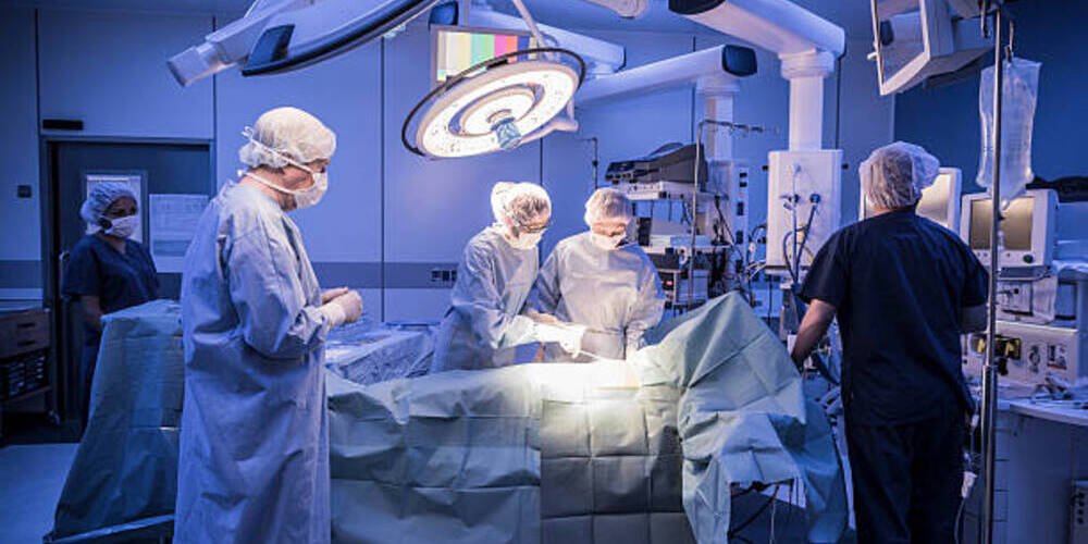
Heart Disease Treatment: Heart Surgery at a Glance
Heart surgery is one of the important ways to treat heart disease. Doctors can perform heart transplantation, implant a rhythm regulator or repair heart valves through surgery. For example, coronary artery bypass surgery is one of the more well-known heart surgeries. .
In recent years, due to the rapid development of minimally invasive surgery, cardiac surgery is no longer a major surgery that requires sawing through the sternum. In comparison, open-heart surgery performed through minimally invasive surgery has less risks for the patient and is more convenient. You can choose the treatment method that suits you.
However, there are many types of cardiac surgeries, and many different names may refer to the same surgery, which often makes people look confused. This article summarizes the various types of cardiac surgeries for you and briefly introduces their uses, differences, risks and Implementation modalities.
Heart coronary surgery
Atherosclerosis in the coronary arteries is often the main reason for coronary artery surgery; if the accumulation of fatty plaque in the arteries is very serious, the patient is likely to suffer from myocardial infarction. Whether a myocardial infarction has occurred or has not yet occurred, patients may need surgery to remove plaque or unblock blood flow. The following is an introduction to common coronary artery surgeries:
Coronary artery bypass surgery
Coronary artery bypass graft (CABG) surgery is performed by transplanting blood vessels elsewhere to bypass blocked coronary arteries and allow blood to circulate. It is mainly used to treat patients with atherosclerosis, angina pectoris and coronary heart disease (also known as ischemic heart disease).
In the past, heart bypass surgery could only be performed through a thoracotomy and carried higher risks, so patients were usually only considered for this procedure if they had several narrowed coronary arteries. However, current technology can now perform minimally invasive robotic arm surgery through the gap between the ribs, giving high-risk patients a more suitable option.
In addition, there is another type of surgery called off-pump coronary artery bypass (OPCAB), which means the surgery is performed while the heart keeps beating without stopping the heart and using an extracorporeal circulation machine. The advantage is that the patient avoids the risk of temporary cardiac arrest, but the procedure is also more difficult to perform.
Percutaneous coronary intervention
Percutaneous coronary intervention, hereinafter referred to as PCI. This procedure is also known by many other names, including Angioplasty, Balloon Angioplasty, Cardiac Catheterization, etc.
The operation involves inserting a catheter through a blood vessel in the groin or arm and passing it through the blood vessel to the heart. A balloon is then sent through the catheter to the narrowed part of the artery. When the balloon is inflated, it can open the blood vessel wall and increase blood flow. In order to prevent the artery from narrowing again after surgery, cardiac angioplasty is usually accompanied by a stent, which is left in the artery that has been opened by a balloon to prevent the wall from retracting.
Atherectomy
Atherosclerotic plaque resection, also known as Atherectomy in English, also uses the method of inserting a catheter. A rotating blade is installed on the top of the catheter to enter the blocked blood vessel to grind and remove the plaque. This procedure may also be used in patients who have had angioplasty but still have plaque clogging their blood vessels.
Heart valve surgery
Heart valves are valves that control blood circulation in the heart. The valves must open and close accurately to prevent blood from flowing backward. However, if you suffer from rheumatic heart disease or congenital heart disease, it may cause valve prolapse and insufficiency, such as the commonly heard mitral valve (mitral valve) prolapse, aortic valve stenosis, etc. At this time, the valve needs to be repaired or replaced to maintain normal heart function. Valve surgery can be divided into repair and replacement. Please see the following introduction: (Extended reading: Valvular heart disease)

Valve repair surgery
In cases where the valve can be repaired, valve repair surgery is often performed to restore valve function. The traditional method is to saw open the sternum and cut into the heart to directly repair the problematic valve. Now, with the help of minimally invasive surgery, such as using the Da Vinci arm, or using video-assisted thoracoscopic surgery (VATS), surgery can be performed from the gap between the ribs without incision. The patient’s ribs can help reduce postoperative pain and shorten the recovery period.
In the case of valve repair surgery, a valve ring is installed to prevent the valve from loosening and improve the problem of insufficiency. If the valve is stenotic, the valve may be slightly cut to allow more blood to pass through.
Transcatheter aortic valve replacement surgery
Transcatheter aortic valve replacement surgery (referred to as TAVI or TAVR), this surgery is actually the same concept as the PCI surgery mentioned above. The catheter is inserted from the groin or arm, reaches the heart through the blood vessels, and then the catheter is fixed on the stent. The artificial heart valve is directly inserted and there is no need to specifically remove the patient’s original heart valve.
However, this “transcatheter” valve replacement surgery is also suitable for replacing the mitral valve or tricuspid valve, not just the aortic valve. Not only that, transcatheter surgery can also use a catheter to carry a valve clip to repair minor injuries. Valvular atresia is a problem, so whether it is valve replacement or repair, transcatheter surgery can be considered.
Heart failure surgery
1.Ventricular assist device
The installation of a ventricular assist device requires open-heart surgery; the doctor will install a pump on the upper abdominal wall, then connect the catheters from the aorta and ventricle to the pump, and connect the pump to the external ventricular assist device with wires. After completion, the blood in the ventricle will flow along the catheter to the pump, and the blood will be pumped into the aorta through the pump to maintain blood circulation. To put it simply, a ventricular assist device actually plays the role of a ventricle.
To break it down, ventricular assist devices are divided into fixed and portable ones. Fixed ventricular assist devices cannot be carried and the patient can only move around in the ward, while portable ones can be fixed on the patient and do not restrict the patient. action.

2.Intraaortic balloon pump
This type of pump, like the ventricular assist device, is one of the ways to assist the heart in maintaining blood circulation. However, the intra-aortic balloon pump (IABP) is more convenient to install and is also the most commonly used. The treatment method is to use a cardiac catheterization method to insert a balloon-carrying catheter into the femoral artery at the groin and guide it to be placed close to the aortic arch. This balloon can collapse during cardiac systole to reduce the afterload of the heart; it can expand during diastole to push blood back up the aorta and increase coronary blood perfusion.
3.Extracorporeal membrane oxygenation
Extracorporeal membrane oxygenation, or ECMO for short. The biggest difference from a ventricular assist device is that the ventricular assist device also has the function of converting blood into oxygenated blood. It is mainly used for patients with severe heart and lung diseases.
There are two main ways to install a membrane: Veno-Venous (VV) and Veno-Arterial (VA). In the former case, catheters are usually inserted into the femoral vein and internal jugular vein, while in the latter case, the femoral vein and femoral artery are inserted. Both of them drain the blood through a pump (artificial heart), remove carbon dioxide from the blood and heat the blood through an extracorporeal oxygenator (artificial lung), and then return it to the veins or arteries.
However, the disadvantage of Extracorporeal membrane oxygenation is that it cannot be used for a long time. The artificial material pipeline and closed circulation system of Extracorporeal membrane oxygenation make patients prone to infection due to hemolysis and blood clots. The ends of hands and feet may gradually turn black and necrotic, and even require amputation.

4.Heart transplant
Heart transplantation is usually performed on patients with end-stage heart failure, when the degree of damage to the heart cannot be improved by medication or any other means. Heart transplantation can only be achieved through open-heart surgery, in which the sternum is sawed open and the old heart is removed and replaced with a healthy heart from a donor.
However, it is not that easy to wait until someone can donate a suitable heart. During the transition period before a heart transplant, patients with heart failure can use a ventricular assist device (VAD) to help their weakening heart pump blood.
5.Total artificial heart
A total artificial heart is a very expensive and sophisticated device. It is different from a ventricular assist device or a ventricular membrane. It needs to have all the functions of a natural heart, including valve design, left and right ventricle and atrium design, etc. In addition, using a total artificial heart means that the patient’s own heart needs to be removed, so the risk is higher. Unless they are patients with advanced heart failure who have both ventricular failure, in most cases only an auxiliary artificial heart or a ventricular assist device will be used as a transitional period for heart transplantation.
Arrhythmia surgery
1.Cardiac catheter radiofrequency cauterization
Radiofrequency catheter ablation (RFCA) in English is mainly used to treat arrhythmia (Arrhythmia), such as atrial fibrillation and paroxysmal supraventricular tachycardia (PSVT).
The operating principle is the same as that of cardiac catheterization. A catheter with an electrode on the tip is inserted into the groin and followed along the blood vessels to the location of the myocardium emitting abnormal heart rhythm. The electrode emits radiofrequency energy to destroy the diseased myocardial cells and return the heart rhythm to normal. In addition to radiofrequency cautery, this surgery also uses cryoablation. The advantage is that the operation time is shorter, but the surgical deductible is higher.
2.Maze surgery
Known as Maze surgery in English, maze surgery uses electrosurgery (or electrocautery) to create scars on the myocardial tissue and change the flow direction of the heart’s electrical circuit. Because scars cannot conduct electricity, they are like walls in a maze, blocking pulmonary veins and Abnormal conduction of electrical current between the atria. The maze procedure is primarily used to treat atrial fibrillation and may also be performed in conjunction with RFCA. (Recommended reading: Maze surgery: Applicable groups, risk factors, surgical procedures and postoperative recovery)
3.Install a heart rate regulator and defibrillator
Some patients with arrhythmias who cannot control their condition with drugs may consider installing a pacemaker (Pacemaker) or an Implantable cardioverter defibrillator (ICD). They can detect the patient’s heart rhythm. If there is an abnormal heart rhythm, situation, it will send out electric current to correct the heartbeat rhythm or perform cardiac defibrillation.
When installing a pacemaker and defibrillator, you usually choose to cut a five-centimeter opening in the patient’s left chest and below the clavicle, bury the machine and venipuncture the wires to connect the pacemaker to the heart. .


